Loculated Pleural Effusion Cxr / Helpful radiological signs in cxr25 11-91 / Pfa = pleural fluid analysis.. It has many causes ( pneumonia, heart failure, blood clots, trauma, bleeding). The first step in evaluating pleural effusions is determining whether it is transudative or exudative. Analyses of the pleural aspirate confirmed a complicated parapneumonic effusion: What does a westermark sign look like? The pleura are thin membranes that line the lungs and the inside of the chest cavity and act to lubricate and facilitate breathing.
Complex septated, complex nonseptated, or homogeneously echogenic effusions are always exudates (fig. It has many causes ( pneumonia, heart failure, blood clots, trauma, bleeding). Weight loss 15 lbs in one month • pf is a transudate; It results when the production of pleural fluid exceeds the body's ability to reabsorb it. Loss of right diaphragmatic and cardiac silhouettes.
Cureus | Rhabdomyolysis as a Presentation of 2019 Novel ... from assets.cureus.com Surgical thoracostomy tube placement and radiologically guided catheter drainage are standard therapy for loculated pleural fluid collections. Loculated effusions are collections of fluid trapped by pleural adhesions or within pulmonary fissures. What are loculated pleural effusions? Fixing the underlying cause with or withourt draining the fluid usually results in cure. What are the different appearances of pleural effusion? An anechoic effusion can be a transudate or exudate (fig. • in patients with symptomatic mpes with nonexpandable lung, failed pleurodesis, or loculated effusion, ipcs are suggested over chemical pleurodesis. The first step in evaluating pleural effusions is determining whether it is transudative or exudative.
It has many causes ( pneumonia, heart failure, blood clots, trauma, bleeding).
Pleural fluid trapped within adhesions and may mimic a nodule, atelectasis, or consolidation often located within fissues. There is a large left pleural effusion obscuring the lower half of the left hemi thorax. It has many causes ( pneumonia, heart failure, blood clots, trauma, bleeding). A chest tube (12f) was inserted under imaging guidance into the largest locule. A repeat cxr and ultrasound on day (d) 5 identified a multiloculated pleural effusion. Differential diagnosis of pleural effusion; But, catheter removal is suggested if the infection fails to improve. Most pleural effusions, whether free flowing or loculated, are hypoechoic with a sharp echogenic line that delineates the visceral pleura and lung. Fluid gathers in the lowest part of the chest, according to the patient's position. All patients were subjected to routine chest radiography (cxr; Ph 6.09, lactate dehydrogenase 71,300 u/l, protein 40 g/l but no microorganism was cultured. It detects pleural effusions with higher sensitivity and specificity than cxr, and provides valuable information about the size and depth of the pleural effusion, the echogenicity of the fluid, the presence of septated or loculated fluid, pleural thickening and nodularity, and the presence of any. It detects pleural effusions with higher sensitivity and specificity than cxr, and provides valuable information about the size and depth of the pleural effusion, the echogenicity of the fluid, the presence of septated or loculated fluid, pleural thickening and nodularity, and the presence of any contralateral pleural effusion.
It results when the production of pleural fluid exceeds the body's ability to reabsorb it. Sometimes in the setting of pleuritis, loculation of fluid may occur within the fissures or between the pleural layers (visceral and parietal). Analyses of the pleural aspirate confirmed a complicated parapneumonic effusion: It detects pleural effusions with higher sensitivity and specificity than cxr, and provides valuable information about the size and depth of the pleural effusion, the echogenicity of the fluid, the presence of septated or loculated fluid, pleural thickening and nodularity, and the presence of any. Pleural effusion is not a disease.
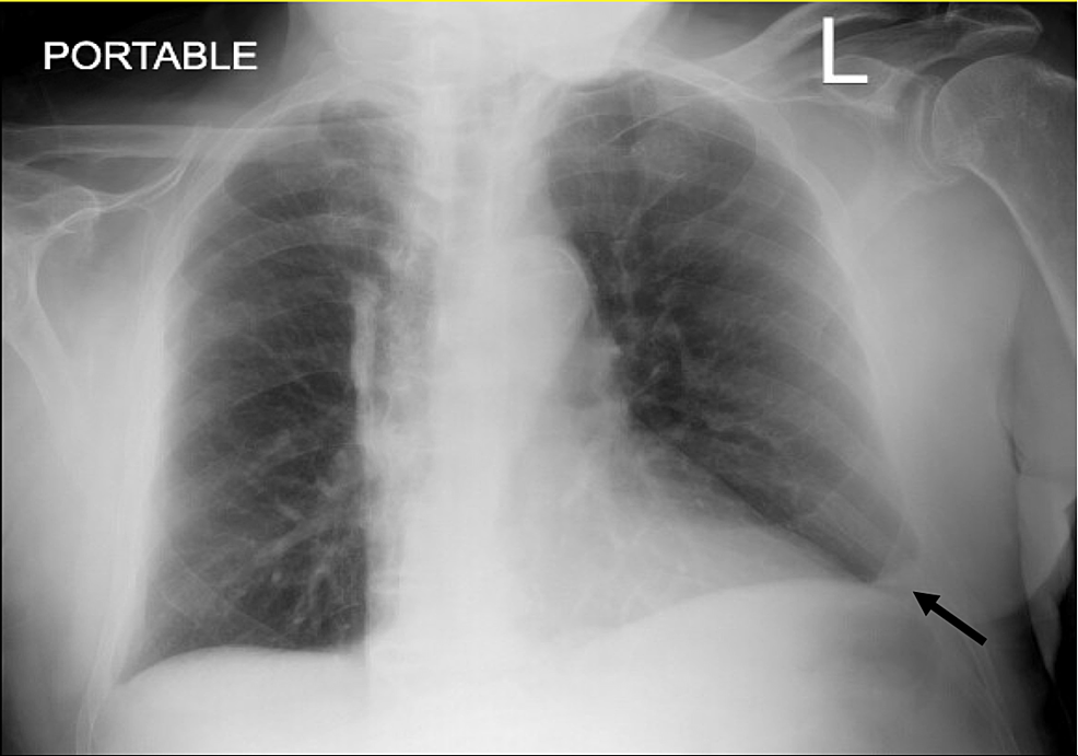
CXR Case 094 • LITFL • Chest X-ray Self-Assessment Quiz from litfl.com There is a large left pleural effusion obscuring the lower half of the left hemi thorax. Transudative effusions are a result of pressure filtration without capillary injury (i.e hydrostatic and oncotic pressure abnormalities). Ask learners to list mechanisms of complex pleural fluid drainage (click on this). An anechoic effusion can be a transudate or exudate (fig. Case 1 • 77 year old woman with hx of copd • 2 week history of uri symptoms • zpak and then 10 days antibiotics • hospitalized with 3 day history of fever to 39.0 °c, shaking chills, nausea and large pleural effusion. Normally, a small amount of fluid is present in the pleura. Ph 6.09, lactate dehydrogenase 71,300 u/l, protein 40 g/l but no microorganism was cultured. Enlarged mediastinal lymph nodes, possibly reactive.
Ask learners to list mechanisms of complex pleural fluid drainage (click on this).
Enlarged mediastinal lymph nodes, possibly reactive. Case 1 • 77 year old woman with hx of copd • 2 week history of uri symptoms • zpak and then 10 days antibiotics • hospitalized with 3 day history of fever to 39.0 °c, shaking chills, nausea and large pleural effusion. The differential diagnosis of loculated pleural effusions within the fissure includes the following: Most pleural effusions, whether free flowing or loculated, are hypoechoic with a sharp echogenic line that delineates the visceral pleura and lung. It detects pleural effusions with higher sensitivity and specificity than cxr, and provides valuable information about the size and depth of the pleural effusion, the echogenicity of the fluid, the presence of septated or loculated fluid, pleural thickening and nodularity, and the presence of any contralateral pleural effusion. The right pe was larger and loculated (by ultrasound). Ray, and after treatment (ie drainage), there should be a difference, however, if a cxr is taken day/ month. Transudative effusions are a result of pressure filtration without capillary injury (i.e hydrostatic and oncotic pressure abnormalities). Cxr = chest x ray; Differential diagnosis of pleural effusion; Fluid gathers in the lowest part of the chest, according to the patient's position. On ct, 21% of pleural effusions showed loculation. Malignant pleural effusion, breast carcinoma, maliganancy:
Loss of right diaphragmatic and cardiac silhouettes. • in patients with symptomatic mpes with nonexpandable lung, failed pleurodesis, or loculated effusion, ipcs are suggested over chemical pleurodesis. Transudative effusions are a result of pressure filtration without capillary injury (i.e hydrostatic and oncotic pressure abnormalities). Pleural fluid glucose < 60 mg/dl; Line not corresponding to fissure.
Chest X-ray showing bilateral pleural effusion. | Download ... from www.researchgate.net Most effusions start like this and can be easily missed. Loculated right pleural effusion with foci of atelectasis and consolidative changes concerning for pneumonia. Cxr = chest x ray; Many factors influence the radiographic findings of pleural effusion, including the nature of the fluid (free vs loculated), the amount of fluid, the patient's position, the radiographic projection, and the presence of underlying lung abnormalities. Pleural fluid trapped within adhesions and may mimic a nodule, atelectasis, or consolidation often located within fissues. It has many causes ( pneumonia, heart failure, blood clots, trauma, bleeding). All patients were subjected to routine chest radiography (cxr; Loculated effusions are collections of fluid trapped by pleural adhesions or within pulmonary fissures.
Pleural effusion is an abnormal accumulation of fluid in the pleural space.
In chf effusions are bilateral and more on right. Pleural effusion is not a disease. Treatment may fail if the catheter is not placed optimally within the loculation or if the fluid is hemorrhagic or fibrinous. Ph 6.09, lactate dehydrogenase 71,300 u/l, protein 40 g/l but no microorganism was cultured. Pleural effusions were observed in 32% and 47% of patients by cxr and ct, respectively. It results when the production of pleural fluid exceeds the body's ability to reabsorb it. A ct study revealed this to be a loculated pleural effusion. Bilateral pleural effusion (bpe) is not an uncommon finding in clinical practice. It detects pleural effusions with higher sensitivity and specificity than cxr, and provides valuable information about the size and depth of the pleural effusion, the echogenicity of the fluid, the presence of septated or loculated fluid, pleural thickening and nodularity, and the presence of any. So pleural effusion is seen on a chest x. Typically, pleural effusions were small (90% occupied less than one third of the hemithorax) and unilateral (85%), but occasionally they reached more than a half of the hemithorax. Analyses of the pleural aspirate confirmed a complicated parapneumonic effusion: A right thoracentesis was performed, and on seeing the biochemistry results, the left side was also punctured.
Most pleural effusions, whether free flowing or loculated, are hypoechoic with a sharp echogenic line that delineates the visceral pleura and lung loculated pleural effusion. But, catheter removal is suggested if the infection fails to improve.
Boedapest Kaart Europa / Boedapest Wikipedia - Boedapest is één van de meest onderschatte steden van europa! . Antieke kaart van europa of een europees land gemaakt door winkler prins, kuyper of richard andree te koop bij voor uw antieke kaart en plattegrond kaarten europa. Boedapest kaart europa (hongarije) te downloaden. Breng je stedentrip in boedapest door met de onderstaande bezienswaardigheden. Boedapest is de hoofdstad van hongarije. Air europa logo sky team logo. Boedapest kent een rijke geschiedenis waar verschillende culturen onderdeel van zijn geweest. Kaart van hotels in gebied boedapest: Boedapest is één van de meest onderschatte steden van europa! De stad is gelegen aan weerszijden van de donau. Waar ligt boedapest in hongarije? Illustraties Lineair Beeldresearch from www.lineairbeeldresearch.nl Kies uit 69 prachtige appartementen op de ...
Bathtub Fixed Glass Panel . With millions of unique furniture, décor, and housewares options, we'll help you find the perfect solution for your style and your home. Fixed glass panel in bathtub. Find tempered glass panels in home renovation materials | home renovation materials for sale in 12mm glass panel with no holes designed for railing, pool available sizes: You can use a single sheet or more of glass. A shower lagoon glass bathtub enclosure is one component in a beautiful bathroom renovation. Total 13 shower doors found. A glass warehouse frameless shower door can instantly make your bathroom look bigger and brighter, adding a fresh and modern feel yet having the versatility to complement any bathroom style. View our concise bath panels range at screwfix. Get a fresh perspective for this online shopping industry by. This fixed panel shower screen is designed for the bathtub and resembles a vintage factory window with multiple glass panes, the full divided w...
Song Codes For Roblox Boombox / How Get The Updated Roblox Song Codes For The Latest And Your Favourite Songs Home Roblox Music Codes - 1844348402 (click the button next to the code to copy it) song information: . If you are looking for a fancier option, check out the dual golden duper fly boombox at r$1,000. No matter how different these boomboxes may look, they're the same thing. You can use the comment box at the. To play music in roblox, you need the boombox. There are a couple of options available, the cheapest being the beat up super jank boombox at r$250. Tiktok songs, rap, and more (december 2020) play popular songs with these roblox music codes. To play music in roblox, you need the boombox. Your description is awaiting moderation. Many players are searching for roblox tiktok music codes and this is where you will get all the best ones. What are roblox boombox codes? Roblox S...


Komentar
Posting Komentar